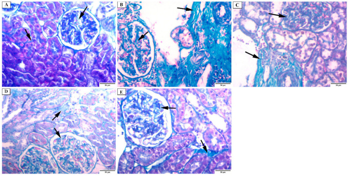
Figure 6.
Photomicrograph of the renal cortex of Masson Trichrome stain showing: (A) group I have a minimal amount of collagen fibers in the renal interstitium and in between the glomerular capillaries (arrows). The basal lamina is positively stained. (B) group II has a massive increase in the collagen fibers in the interstitium and in between the glomerular capillaries (arrows). (C) group III has an intense increase in collagen fibers in the interstitium and between the glomerular capillaries (arrows). (D) group IV has a moderate increase in the collagen fibers in the interstitium and in between the glomerular capillaries (arrows). (E) group V has a mild increase in the collagen fibers in the interstitium and in between the glomerular capillaries (arrows) (×200).

