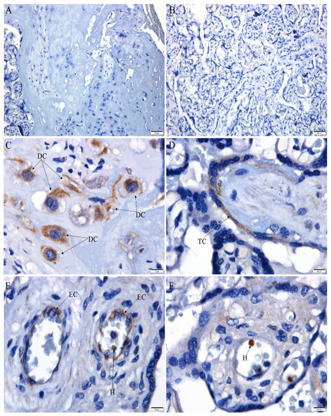
Figure 2.
CHIKV antigen detection in the placenta. (A–B) Negative CHIKV antigen detection in the control placenta; (C–F) infected placenta with CHIKV antigen detection in: (C) decidual cells (DC), (D) trophoblast cells (TC), (E) endothelial cells (EC) and cell inside fetal capillary (H), and (F) cell inside fetal capillary (H). Scale bar—(A,B): 50 µm; (C–F): 10 µm).

