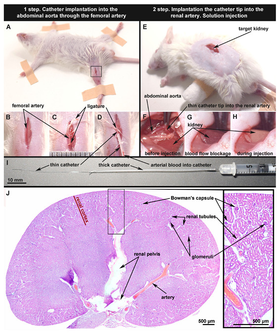
Figure 1.

Main stages of the minimally invasive mouse renal artery catheterization technique (A–H) using polyurethane tubing (I); skin incision on the right thigh for access to the femoral artery marked with a black square (A). Renal artery before (B) and after ligation (C). The thin part of the catheter (4.5 cm, inner diameter 0.25 mm) is completely implanted in the renal artery. The arterial blood in the catheter indicates that implantation was performed correctly (D). Skin incision in parallel with the spine above the kidney for access to the abdominal aorta and renal artery (E). Catheter tip pushed into the renal artery through the abdominal aorta; the artery’s blood flow is preserved, and the targeted kidney has a normal color (F). Renal artery walls are tightly pressed to the catheter walls using tweezers to prevent the entering of injection solution into the aorta; the kidney has a dark color due to the blood flow blockage (G). The kidney color change from dark to light during injection indicates that the manipulation was performed correctly (H). The subgross histological image of the target kidney 5 days after the saline injection (sham operation) shows that the kidney tissue has a normal structure. The thickness of the histological samples is 5 m; dyes: hematoxylin and eosin (H & E) (J).
