
Figure 4.
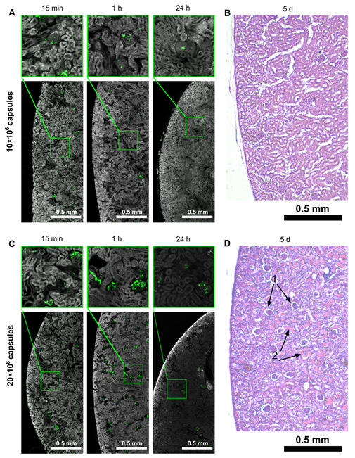
Distribution of capsules administered to the left kidney and dose-dependent impact on the kidney’s structure. Fluorescent microscopy of the target kidney’s cryosections 15 min, 1, and 24 h after microcapsules’ injection at a dose of 10 × 10 (A). Histological section of the target kidney 5 days after 10 × 10 capsules’ administration (H & E staining) (B). Fluorescent microscopy of the target kidney’s cryosections 15 min, 1, and 24 h after microcapsules’ injection at a dose of 20 × 10 (C). Histological section of the target kidney 5 days after 20 × 10 capsules’ administration (H & E staining). 1—Bowman’s capsule is dilated, 2—plasma proteins in the lumen of the tubule (D).