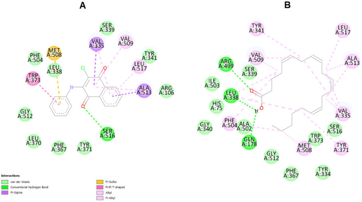
Figure 11.
(A) Visualization of the interactions between COX-2 enzyme and arachidonic acid in their binding sites. Highlighted are the van der Waals interactions (pink) and hydrogen bonds (green). (B) Visualization of the interactions between COX-2 enzyme and compound 9. Note that the binding site is the same as arachidonic acid. In pink: van der Waals interactions; in yellow: π-sulfur interactions; in green: hydrogen bonds (green); purple: π-σ interaction between hydrophobic amino acids (Valine 335 and Alanine 513) and the naphthoquinone moiety of compound 9.

