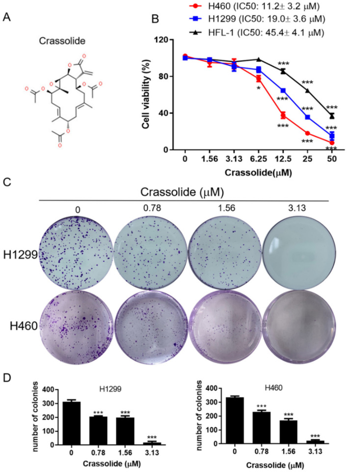
Figure 1.
(A) Chemical structure of crassolide. (B) H460, H1299, and HFL-1 cells were treated with crassolide (0 to 50 μM) for 24 h. The cell viability was detected via an MTT assay. (C) Colony formation assays of H1299 and H460 cells treated with 0–3.13 μM crassolide for 7 days were demonstrated. (D) Data of colony formation assays are presented as mean ± SEM of three wells from one of three experiments. Compared to the DMSO-treated control group, significant differences are indicated by * p < 0.05, and *** p < 0.001.

