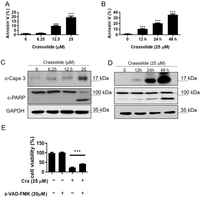
Figure 3.
The effect of crassolide on caspase-dependent apoptosis of H460 cells. (A) H460 cells were treated with different concentrations of crassolide for 24 h or (B) subjected to treatment with 25 µM crassolide for different time points. We measured phosphatidylserine externalization and DNA integrity by FITC-annexin-V and PI, respectively. Annexin-V+/PI− staining (lower-right quadrant) indicates early apoptosis, while Annexin V+/PI+ staining (upper-right quadrant) represents late apoptosis. Data are presented as mean ± SEM of three wells from one of three experiments. Significant differences from the DMSO-treated control group were indicated by *** p < 0.001. The expression of cleaved caspase-3 and PARP was measured by Western blotting after treatment with crassolide at different doses (C) or at different times (D). Data are presented as mean ± SEM (n = 3) for three independent experiments. (E) The viability of H460 cells after treatment with a pan-caspase inhibitor (Z-VAD-FMK) and crassolide. After 24 h, cell proliferation was measured using the MTT assay. Data are presented as mean ± SEM of three wells from one of three experiments, while *** p < 0.001 indicates a significant difference from the group treated with crassolide alone.

