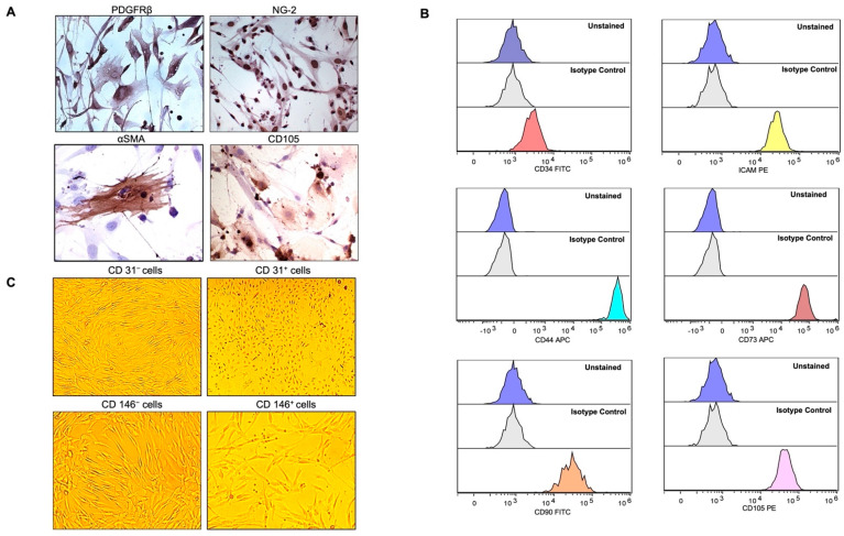
Figure 1.
Expression of mesenchymal cell markers (MSCs) on cultured pericytes (PCs). (A) Immunohistochemistry staining with CD31− PCs were strongly positive for PDGFRβ, NG-2, and most of the cells were positive for CD105 but slightly for αSMA. Pictures were taken at magnification 40× after avidin-biotin peroxide staining. (B) Flow cytometry demonstrated CD31− PCs to be mostly negative for CD34, and positive for ICAM1, CD44, CD73, CD90, and CD105. Histograms in the lowest panels (multicolor) represent the staining for specific antibodies as compared to the unstained and unrelated isotype-matched antibodies in blue and light grey histograms, respectively. (C) Bright-field microscopy depicting the morphology of CD31− PCs versus CD31+ cells as the latter were distinguished by the endothelial-cell-like cobblestone morphology from the elongated and slender shape of the former. CD31− cells, selected for CD146-based MACS (CD146− and CD146+), were similar in shape and morphology. Pictures were taken at a magnification of 10×.

