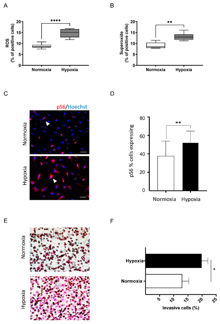
Figure 3.
Cells in the deoxidizing absorbers chamber displayed increased levels of ROS, Superoxide, mTOR levels, and invasive capacity. Cells cultivated in the presence of deoxidizing display (A) increased ROS and (B) superoxide levels (**** p < 0.00001, ** p < 0.001, respectively). (C) Immunofluorescence demonstrates increased expression levels of the mTOR readout marker pS6 in cells cultured under hypoxia (scale bar 50 µm). (D) Quantification of positive cells for pS6 shows a statistically significant activation of mTOR signaling under hypoxia compared to the control (normoxia) (** p< 0.001). (E) Invasion assay using Millicell cell culture inserts during normoxia and hypoxia, demonstrating the enhanced invasive capacity of tumor cells cultured under hypoxic conditions. (F) Quantification of invading cells, demonstrating a statistically significant increase in invasive behavior in tumor cells under hypoxia (* p< 0.05).

