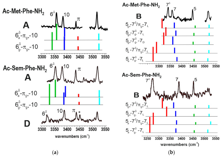
Figure 4.
(a) left panel: IR/UV double resonance spectra of folded conformers observed in the Sem-Phe and Met-Phe sequences, compared to the theoretical spectra (colored sticks) of selected best matching, low energy conformations. The comparison illustrates the similarities of the 6δ H-bonds in these sequences; (b) right panel: same pictures for the semi-extended conformers B, emphasizing the apparent differences in the 7δ H-bonds between the two sequences. See text for assignment.

