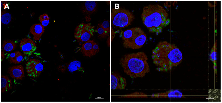
Figure 4.
Spike protein production by engineered L. tarentolae after internalization in the dendritic cells. The presence of the spike protein on the parasite was evaluated by immunofluorescence after 4 h of co-incubation with human dendritic cells. After 4 h, cells were fixed and stained with SARS-CoV-2 spike antibody followed by Alexa Fluor 488-conjugated anti-rabbit IgG secondary antibody. (A) Green dots show the presence of spike protein in the cytoplasm of Leishmania, while the red signal (Nile red staining) shows the cytoplasm of the cells. The nucleus and kinetoplastid DNA was stained with DAPI (blue). (B) A magnification, with an orthogonal projection, showed the co-localization of the parasite and the cytoplasm of the cells as indicated by the presence of yellow/orange signals visible on the axes.

