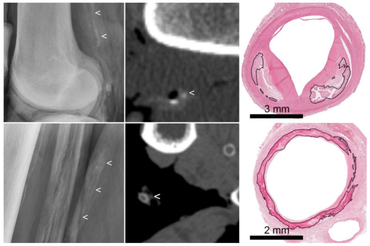
Figure 1.
Examples of intimal and medial calcification on radiograph and CT. (Top row): atherosclerotic intimal calcifications. From left to right: radiograph (showing irregularly distributed thick calcifications (<)), CT (showing thick dots of calcification (<)), and histology (showing calcifications (marked) located in an atherosclerotic plaque). (Bottom row): medial calcifications. From left to right: radiograph (showing regularly distributed thin calcifications along the vascular wall (<)), CT (showing circular thin calcifications (<)), and histology (showing circular calcifications (marked) involving the internal elastic lamina, in the absence of atherosclerosis).

