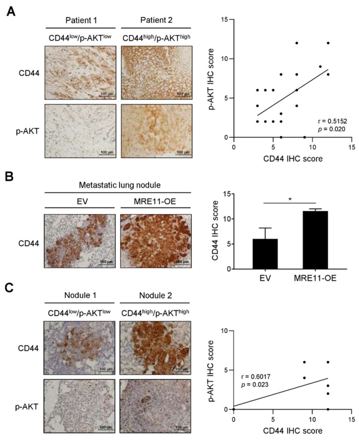
Figure 4.
Correlation analysis for the expression of CD44 and AKT phosphorylation in OSCC patients and metastatic mouse model. (A) Representative images were presented for immunohistochemistry (IHC) staining of CD44 and phosphorylated AKT (p-AKT) expression in the tumor tissue sections from OSCC patients. The quantitative IHC scores were analyzed by Pearson correlation (r) between CD44 and p-AKT (n = 20). (B,C) CAL-27 cells carrying overexpression vector of MRE11 (MRE11-OE) or empty vector (EV) were collected and injected intravenously through the tail vein in mice. The lungs were collected after 8 weeks post-injection for IHC analysis. In (B), representative images were presented for IHC staining of CD44 in the metastatic lung nodules, and the quantitative IHC scores were presented as mean ± SEM (n = 7 per group). Statistical difference was determined by Student’s t-test. * p < 0.05. In (C), representative images were presented for IHC staining of CD44 and p-AKT expression in the metastatic lung nodules from xenograft mice. The quantitative IHC scores were analyzed by Pearson correlation (r) between CD44 and p-AKT (n = 14).

