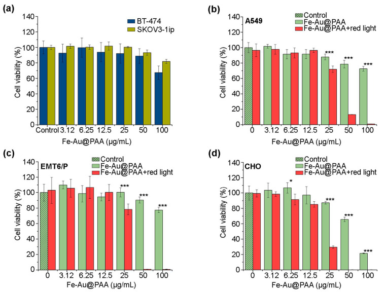
Figure 4.
Analysis of the Fe-Au@PAA nanoparticle cytotoxicity using the MTT test. (a) Study of the toxic effect of particles upon exposure to cells BT-474 and SKOV3-1ip. (b–d) Investigation of the photothermic properties of particles. Cells were incubated with Fe-Au@PAA NPs at various concentrations and irradiated for 7 min with an 808 nm laser at 0.76 W power. Cell viability is shown as a percentage normalized to control cells incubated without particles and irradiation. The statistical differences were considered significant when the p value was < 0.05 (*), 0.001 (***), Welch’s t-test.

