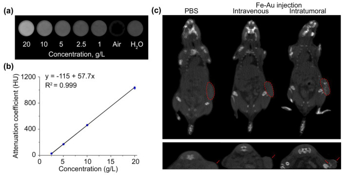
Figure 8.
Application of Fe-Au@PAA NPs as CT contrast agents. (a) CT images of NP solutions at 1–20 g/L concentrations, distilled water, and air. (b) X-ray attenuation coefficient as a function of Fe-Au@PAA NP concentration. (c) CT images of EMT6/P tumor bearing mice before and after intravenous or intratumoral injections of Fe-Au@PAA NPs. The tumor boundary is indicated by a red dashed line in coronal projections (top) or with red arrows at axial projections (down).

