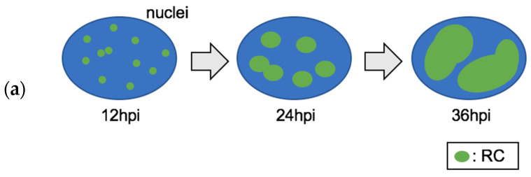
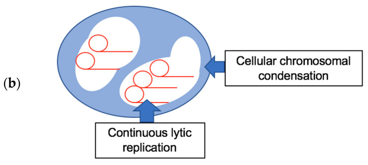
Figure 1.
(a) The formation and growth of RCs over time. These small RCs, seem at 12 hpi, grow bigger and seem to fuse with each other as lytic replication proceeds (24 hpi). At 36 hpi, RCs appear as one or two large globular nuclear subdomains. (b) Model showing the development of RCs and their occupation of extrachromosomal space.


