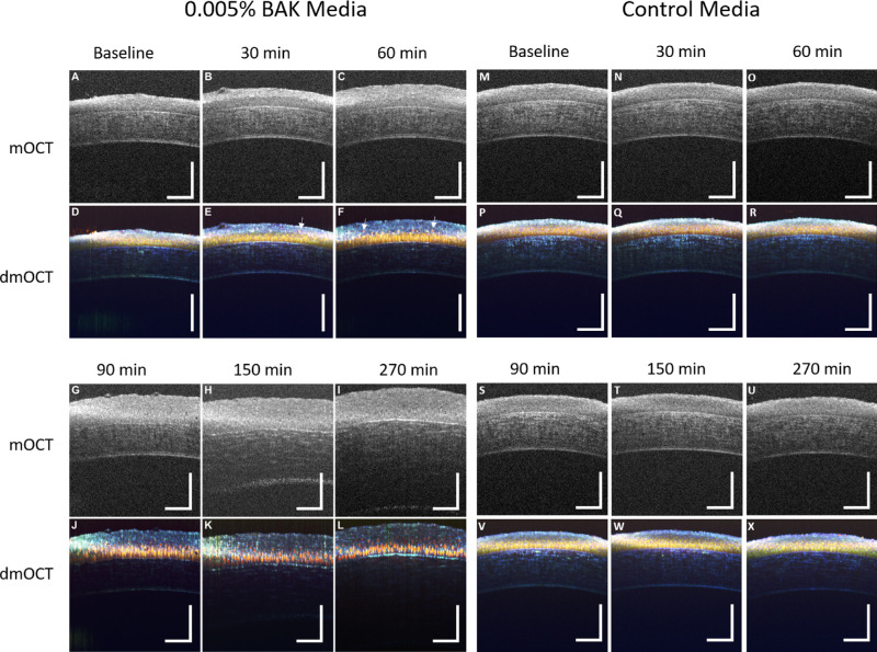
Figure 3.
dmOCT in ex vivo cornea during constant exposure to 0.005% BAK. (A) Single mOCT B-scan in healthy ex vivo cornea prior to exposure to BAK. Scan bar: 100 µm. (B, C) Single mOCT B-scans after 30-minute and 60-minute exposures to 0.005% BAK show swelling of cornea epithelium. (D) dmOCT B-scan in healthy ex vivo cornea prior to exposure to BAK showing superficial and basal epithelium with uniform cell density and tight junctions between basal epithelial cells. (E, F) dmOCT B-scans after 30-minute and 60-minute exposures to 0.005% BAK showing superficial epithelium cells separating from deeper layer and slight swelling of the cornea epithelium. Examples of these cells separating are highlighted with white arrows. (G) Single mOCT B-scan after 90 minutes in 0.005% BAK. (H) Single mOCT scan showing swelling of cornea stroma after 150-minute exposure to 0.005% BAK. (I) Single mOCT B-scan after 270-minute exposure to 0.005% BAK shows severe swelling of stroma. (J) dmOCT B-scan after 90 minutes in 0.005% BAK shows a significant red shift in basal epithelial cells. (K, L) dmOCT B-scans after 120 minutes and 270 minutes in 0.005% BAK show progressive deterioration of cornea epithelium. (M–X) Corresponding mOCT images of healthy ex vivo cornea at different times after immersion in cell culture media. No changes of scattering intensity and dynamic contrast were visible in the control measurements up to 270 minutes.

