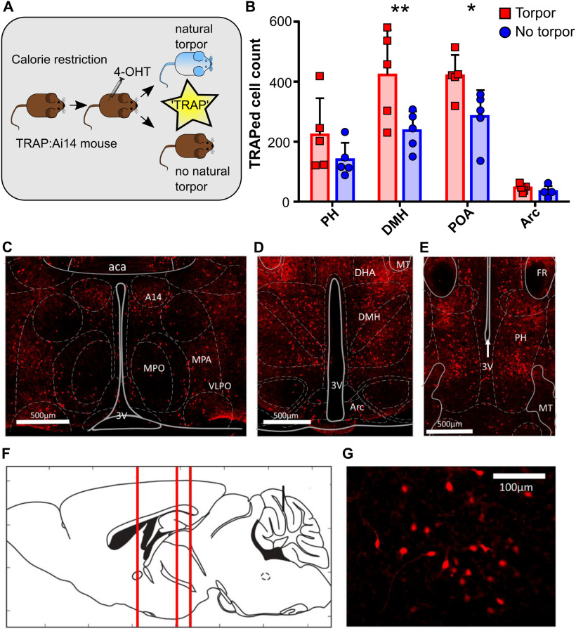
Figure 2.
Torpor is associated with increased activity in dorsomedial and preoptic hypothalamus (n = 5 female TRAP:Ai14 mice). A, Torpor TRAP protocol for TRAP2:Ai14 mice. Calorie-restricted TRAP:Ai14 mice received 4-OHT at ZT7; those that entered torpor following the injection are compared with those that did not. B, Counts of td-Tomato-positive cells by ROI in calorie-restricted mice that entered torpor following 4-OHT (red) or did not enter torpor following 4-OHT (blue). Torpor was associated with higher numbers of TRAPed cells in the DMH and POA compared with animals that did not enter torpor. Repeated-measures ANOVA comparing TRAPed cell count in mice that entered torpor with those that did not for each of the four ROIs. Main effect for torpor versus no torpor (p < 0.05). Main effect for ROI (p < 0.0001). Significant torpor versus no torpor by ROI interaction (p < 0.05). Holm–Sidak's multiple comparisons test: **p < 0.01; *p < 0.05. Data are mean and SD. C, Coronal section showing POA neurons. D, DMH and Arc neurons. E, PH neurons. F, Sagittal schematic showing corresponding anterior-posterior location of coronal sections C–E. G, High-magnification image showing DMH torpor-active neurons expressing td-Tomato. aca, Anterior commissure (anterior part); A14, A14 dopamine cells; Arc, arcuate nucleus; DHA, dorsal hypothalamic area; MPO, medial preoptic nucleus; MPA, medial preoptic area; PH, posterior hypothalamic nucleus; POA, preoptic area; VLPO, ventrolateral preoptic nucleus; 3V, third ventricle. n = 5 female mice per group.

