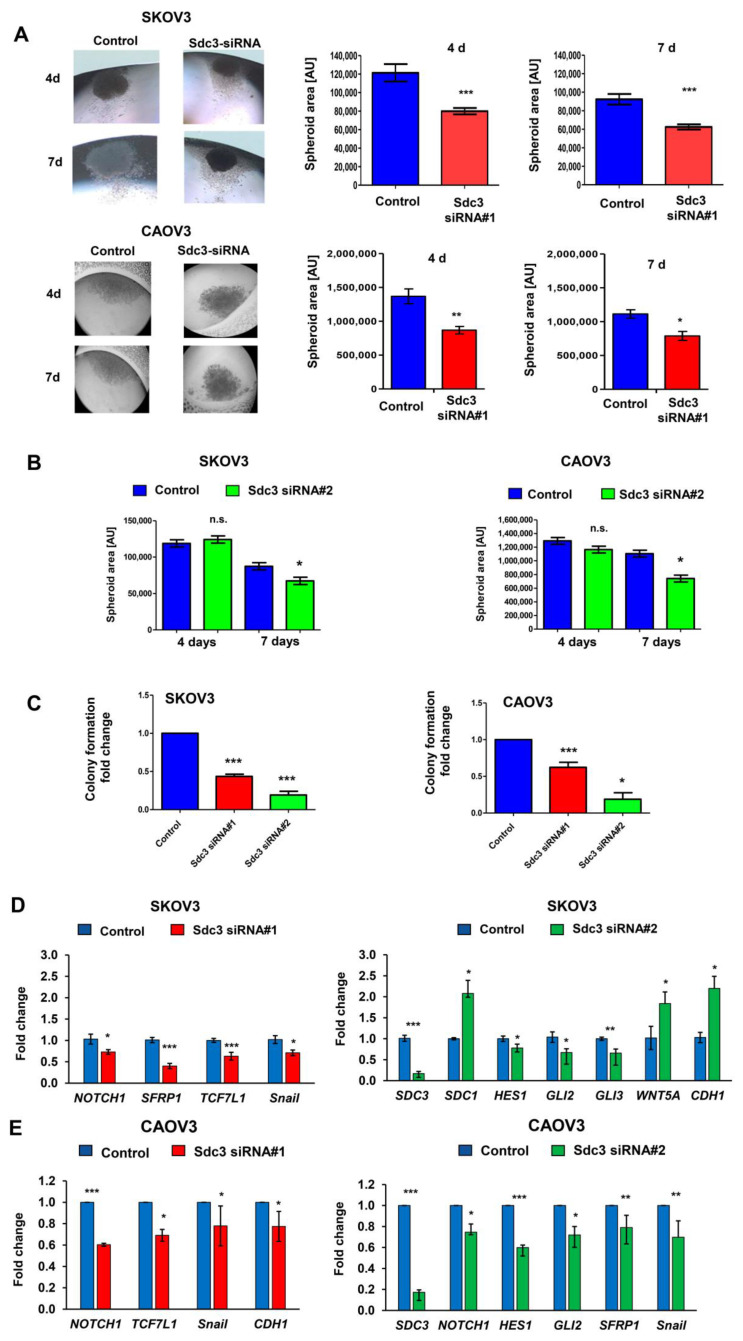
Figure 2.
SDC3 depletion reduces the size of 3D SKOV3 spheroids and alters stemness-related gene expression. (A) Control siRNA and SDC3-siRNA#1- and #2-treated SKOV3 and CAOV3 cells were subjected to a hanging drop assay, allowing for the formation of 3D spheroids [24]. Representative pictures of the hanging drop cultures are presented in the left panels and quantitative analysis in the right panels. The spheroid area was quantified using NIH Image J software. In SDC3-siRNA#1-treated SKOV3 and CAOV3 cells, the spheroid area was significantly reduced after 4 days (p < 0.001, n = 4, error bars = SEM) and 7 days (p < 0.001, n = 4, error bars = SEM) of hanging drop culture, respectively. (B) Quantitative analysis of the spheroid area in SDC3-siRNA#2-treated SKOV3 and CAOV3 cells. The spheroid area was significantly reduced after 7 days (* p < 0.05, error bars = SEM). (C) Colony formation was decreased in SDC3-siRNA#1- and #2-treated SKOV3 and CAOV3 cells. *** = p < 0.001 and * = p < 0.05 error bars = SD. (D,E) qPCR analysis reveals significant mRNA expression changes of constituents of the stemness-related notch and Wnt signaling pathways and of the EMT marker Snail in SKOV3 (D) and CAOV3 (E) after knockdown of SDC3 with the two siRNAs. *** = p < 0.001, ** = p < 0.01, * = p < 0.05, n = 4, error bars = SEM.

