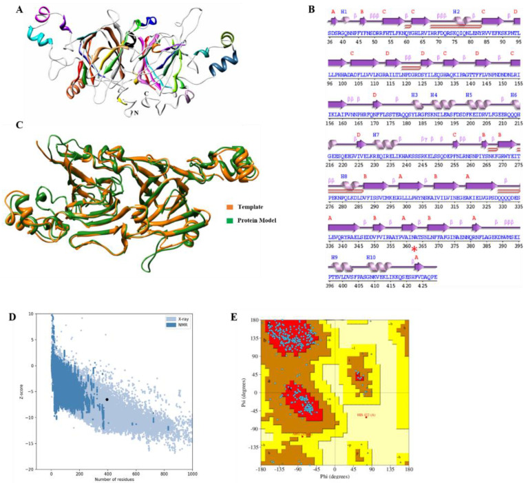
Figure 4.
3D-structure in VacV protein. (A) Ribbon representation of Vicilin protein three-dimensional model, different components of protein such as helices, sheets and loops are highlighted with different colors. N-terminus and C-terminus of protein are shown with N and C, respectively. (B) Secondary structure of VacV protein. (C) Superimposition of vicilin template and globulin protein, Green; Vicilin protein model, and gold globulin protein model. (D) Z-score calculation to estimate the quality of model; black dot representing the position of predicted model. (E) Ramachandran plot; blue dots showing amino acid residues. Upper left and lower left panel belong to allowed regions, while upper right and lower right are the disallowed regions.

