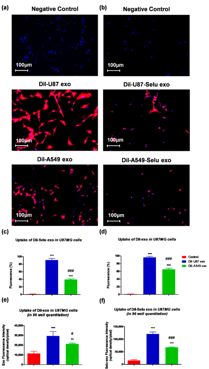
Figure 6.
In vitro exosome targeting to U87 cells. (a,c) DiI-U87 exo showed higher fluorescence intensity than DiI-A549 exo in U87 cells. *** p < 0.001 vs. non-treated control, ### p < 0.001 vs. DiI-A549-Selu exo. n = 3. (b,d) DiI-U87-Selu exo showed higher fluorescence intensity than DiI-A549-Selu exo in U87 cells. *** p < 0.001 vs. control, ### p < 0.001 vs. DiI-A549-Selu exo. n = 3. (e,f) In the 96-well plate, the quantitative uptake of DiI-U87 exo and DiI-U87-Selu exo were higher than DiI-A549 exo and DiI-A549-Selu exo. ** p < 0.01, *** p < 0.001 vs. non-treated control; # p < 0.05, ### p < 0.001 vs. DiI-A549-Selu exo. n = 8. Data are represented as mean ± SD.

