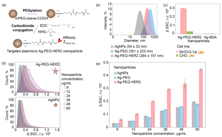
Figure 4.
Nanoparticle conjugation to HER2-recognizing scaffold protein ZHER2:342 and HER2-overexpressing cell targeting in in vitro tests. (a): The schematic illustration of NP surface coating with artificial scaffold polypeptide—affibody ZHER2:342 through the intermediate PEGylation and subsequent carbodiimide conjugation. (b): DLS distributions showing the dependence of the intensity (%) of pristine (Ag NPs), PEGylated (Ag-PEG), and affibody-decorated (Ag-PEG-HER2) silver nanoparticles on their hydrodynamic diameter (nm). (c): Flow cytometry side scatter change ΔSSC of SKOV3-1ip and CHO cell populations labeled with targeted Ag-PEG-HER2 and nontargeted Ag-BSA nanoparticles (r.u.·105). (d): Flow cytometry histograms in SSC channel of SKOV3-1ip cells labeled with different concentrations of Ag NPs (top) and Ag-PEG-HER2 (bottom) nanoparticles. (e) The dependence of ΔSSC (r.u.·103) on SKOV3-1ip cell populations labeled with different concentrations of Ag-PEG-HER2, Ag-PEG, and Ag (µg/mL) obtained with the flow cytometry. ΔSSC was calculated as raw SSC intensity with subtracted SSC intensity of unstained cell samples.

