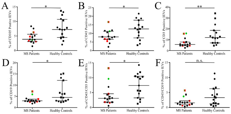
Figure 5.
Relative number of labelled isolated lEVs in MS patients and HC. Percentage of endothelial CD105+ (A), leukocyte CD45+ (B), T-lymphocyte CD3+ (C) and CD45+CD3+ (E), B-lymphocyte CD19+ (D) and CD45+CD19+ (F) lEVs out of all events collected in lEVs gate. The line represents median value with interquartile range. Women (circles), men (squares), patients without treatment (green), patients receiving intravenous corticoids up to 14 days before blood collection (red). * p < 0.05, ** p < 0.005, n.s.—not significant; MS patients (n = 15) and HC (n = 15 or n = 16).

