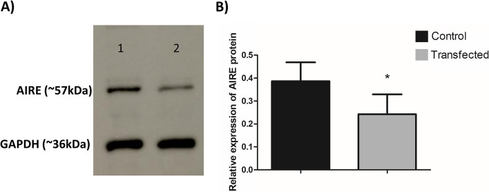
Fig. 5.
Expression of AIRE protein in mTEC 3.10 cell line as detected by western-blot. The mTEC cells were transfected (or not) with miR-155-5p mimic, causing significant decrease in the levels of this protein. A western-blot detection of AIRE after 24 h of transfection. The GAPDH protein was used as housekeeping and to normalize data. The lanes/bands showed were cropped from the blot displayed in the Supp. Figure 1. B Bar graph resulted from quantification of western-blot bands show the effect of miR-155-5p mimic transfection on AIRE expression. The data are presented as the means and standard error of mean (SEM) from three independent determinations. Difference between groups was analyzed by Student t-test, comparing control (not transfected) versus transfected cells (**P < 0.01)

