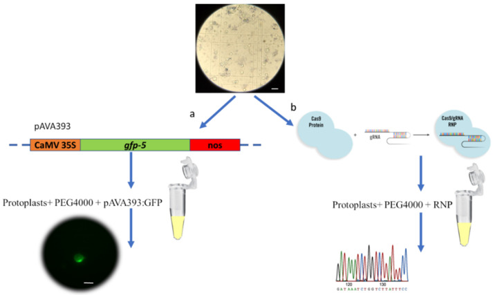
Figure 5.
PEG-mediated transfection protocol. (a) Protoplast transfection using GFP marker gene and subsequent visualization using the Nikon Eclipse Ti2 fluorescent microscope. (b) Protoplast transfection using RNP complex by targeting the pds gene, followed by DNA extraction and Sanger sequencing. Scale bar = 100 µm.

