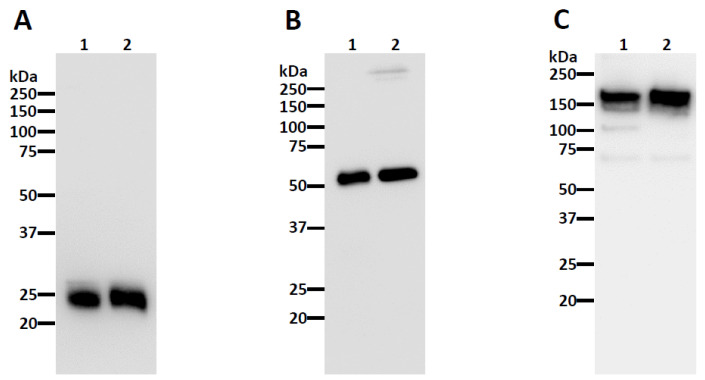
Figure 2.
Western blot analysis of plant-made CA1. Plant-made CA1 was subjected to SDS-PAGE under reducing conditions (A,B) and non-reducing conditions (C). Proteins were transferred to a PVDF membrane after separation and a horseradish peroxidase-conjugated goat anti-human kappa (A,C) or goat anti-human IgG (B) antibody was used to detect the light chain and heavy chain, respectively. Lane 1, plant-made CA1; Lane 2, mammalian cell-produced anti-West Nile virus E protein (E16) IgG. The blots are representatives of multiple independent experiments.

