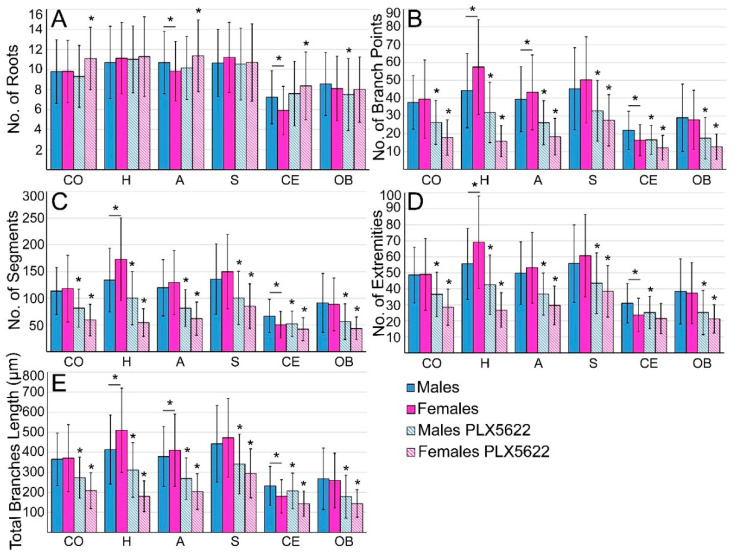
Figure 4.
Quantitative morphological analysis of microglia in different brain regions in control male and female rats and rats fed ad libitum for ten days on PLX5622 chow. Controls (homogeneous columns, male in blue and female in magenta), PLX5622-treated rats (in diagonal stripes). (A) The number of roots/cell, (B) number of branching points, (C) number of segments, (D) number of extremities, and (E) total branch lengths. The number of roots/microglia in control and microglia-surviving PLX5622 in the cerebellum and olfactory bulb was lower than in other brain regions and was similar in males and females in most of the brain regions that were examined and was not altered by PLX5622 treatment. Microglia surviving the 10 days of PLX5622 chow showed a reduced number of branching points, segments, extremities, and total branch lengths in all examined brain regions. CO—cortex, H—hippocampus, A—amygdala, S—striatum, CE—cerebellum, OB—olfactory bulb. Data are presented as mean ± one standard deviation. ≥138 cells were analyzed from slices prepared from the six different areas of interest of the different hemispheres (4 controls and 4 PLX5622-fed rats) of each sex. Significant differences were determined by a t-test for two samples assuming unequal variances. p value ≤ 0.01 indicated a statistically significant difference. Underlined asterisks indicate statistical significance between males and females; asterisks indicate statistical significance between control and PLX5622-fed rats; vertical lines correspond to ± one standard deviation.

