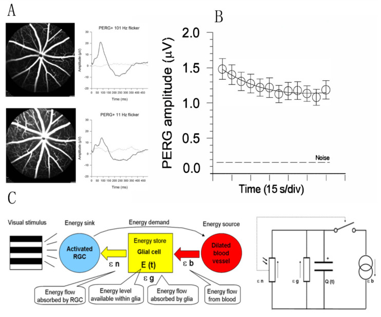
Figure 6.
Flicker-induced PERG adaptation. (A) Flickering light at 11 Hz superimposed to a pattern stimulus induces vasodilation in C57BL/6J mouse, as shown by fluorescein angiography, and reduces the PERG amplitude compared to the same pattern stimulus with superimposed flicker at 101 Hz (invisible to photoreceptors). (B) In human subjects, the PERG signal in response to sustained pattern reversal at 16 rps becomes progressively reduced to a plateau over 2 min. (C) Energy budget model that accounts for the temporal dynamics of PERG adaptation in mice and human subjects. At any given time, the energy available to activated neurons (ε n, photoresistor) depends on the energy flow provided by glial stores (ε g capacitor) and vascular supply (ε b, current generator) minus the energy absorbed in the process (ε g, resistor). The switch connecting activated neurons to vascular supply represents the neurovascular coupling. The direction of arrows indicates the energy flow. (A) Replotted from Chou et al., Sci. Rep. 2019; (B) replotted from Porciatti et al., IOVS 2005; (C) replotted from Porciatti and Ventura, Vis. Res. 2009.

