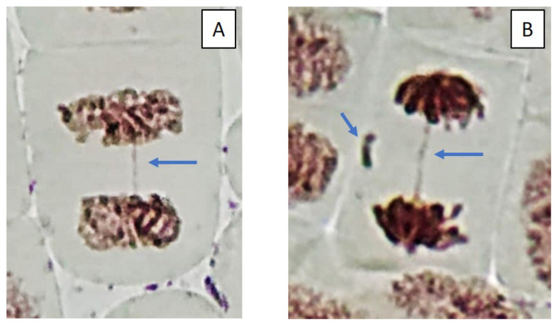
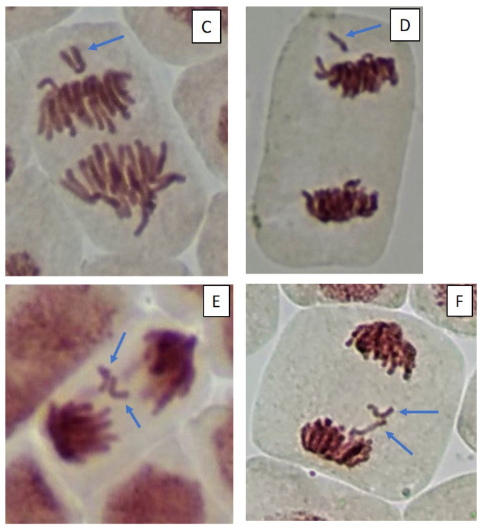
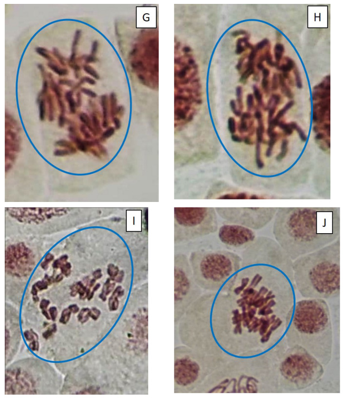
Figure 5.
Microphotography of cells in the root tips of Allium cepa L. exposed to the water from pools with model microcosms. Location of chromosome aberrations in the cells indicated by arrows. Mitotic abnormalities and polyploidy indicated by circles. Types of abnormalities: (A) chromosome bridge; (B) chromosome bridge and single fragment; (C) vagrant chromosome; (D) fragment; (E) two lagging chromosomes; (F) lagging chromosome and fragment; (G) mitotic spindle disturbances; (H) mitotic spindle disturbances; (I) C-mitosis; (J) C-mitosis; (K) polyploidy; (L) polyploidy with lagging chromosomes; (M) small micronuclei (formation from fragments); (N) large single micronuclei (formed from lagging chromosome).


