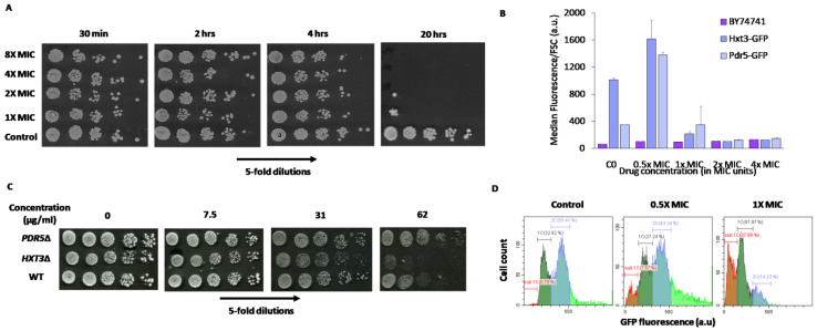
Figure 5.
Effects of Mycosidine on cell growth and death and levels of several GFP-tagged proteins in S. cerevisiae yeast. (A) Spotting of BY4741 yeast treated with Mycosidine onto YPD shows that the drug efficiently kills yeast cells but only after a 24-h incubation; (B) Flow-cytometric analysis of GFP-fusion protein fluorescence in yeast cells shows that Pdr5 and Hxt3 are induced after 0.5×MIC (7.8 mg/L) treatment of S. cerevisiae cells by Mycosidine. Presented data are median fluorescence values for living cells (negative for PI staining); (C) Yeast strains (BY4742) with deletions of the noted genes were spotted onto YPD plates containing the noted concentrations of Mycosidine. Deletion of the Pdr5 gene did not cause increased sensitivity to Mycosidine, whereas deletion of HXT3 did; (D) Distributions of Htb2-GFP fluorescence in live cells treated with different concentrations of Mycosidine. Sub1C (orange), 1C (green), and 2C (grey) denote the putative amount of DNA in the cells. The percentage of cells in each population is noted.

