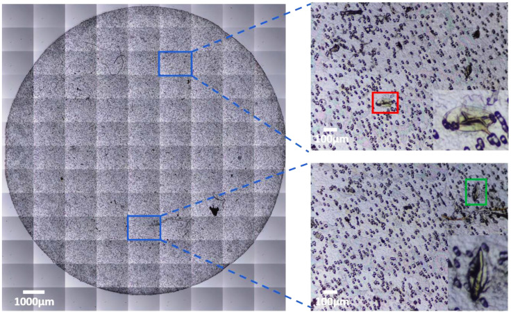
Figure 2.
Automated image grid acquisition of Schistosoma eggs from a urine filter membrane. Blue region of interest shows individual sub-images, red and green regions of interest are S. mansoni and S. haematobium eggs, respectively, present in the urine sample. Enlarged areas show the eggs at 300% digital zoom.

