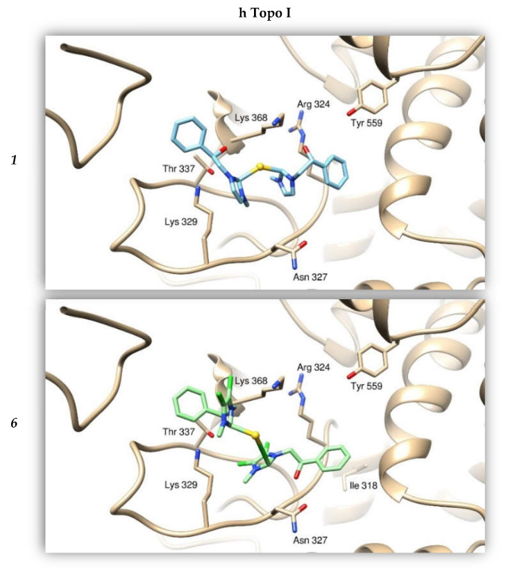
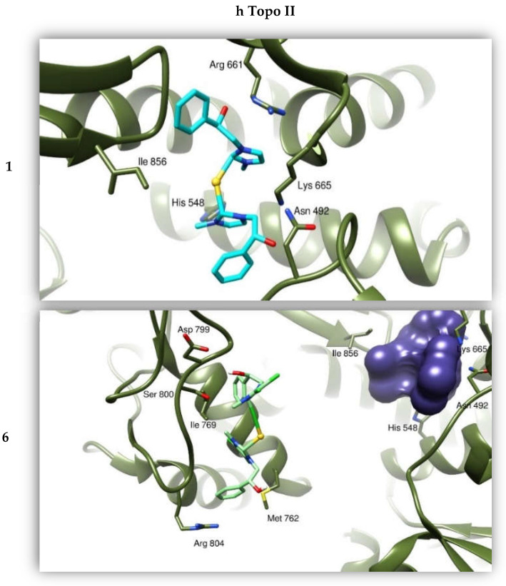
Figure 7.
Binding modes of compounds 1 and 6 with hTopo I and hTopo II. The proteins are depicted as tanned and olive-green ribbons, respectively. Compound 1 is drawn as light blue sticks, while compound 6 as green sticks. As reference, the violet bubble indicates the position of compound 1 with respect to compound 6 in binding hTopo II.


