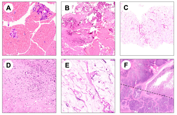
Figure 1.
Representative images of potential pitfalls in NGS analysis of FFPE bioptic and surgical specimens. (A,B) Hematic material enclosing few adenocarcinoma glands; (C) biopsy specimen composed of fibrotic tissue enclosing rare adenocarcinoma glands; (D) scattered tumor cells surrounded by necrosis and fibrosis in a metastatic surgical resection specimen; (E) mucinous adenocarcinoma characterized by low cellularity and mucinous acellular component; (F) intratumor heterogeneity may hamper NGS analysis if both components are not considered (a morphologically heterogeneous colorectal adenocarcinoma composed by glandular and solid areas is shown).

