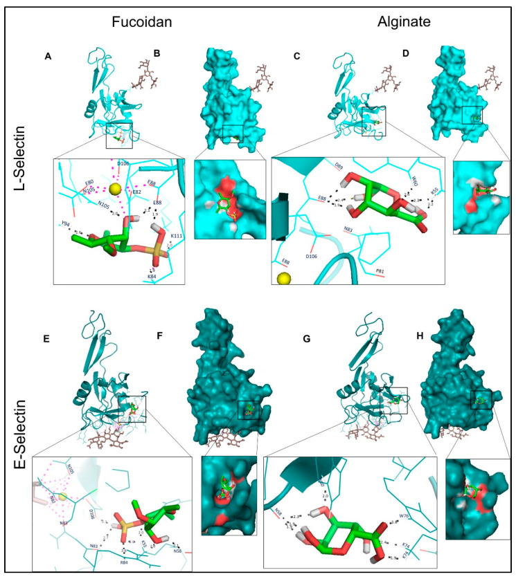
Figure 4.
Molecular docking of L-and E-selectins with fucoidan and alginate. (A,C,E,G): 3D structures of proteins bound with N-linked glycan moieties (brown) and zoomed ligand-binding pocket. Black and purple dotted lines, respectively, describe the H-bonds and Ca2+ (yellow ball) coordination bonds. (B,D,F,H): molecular surface representation, and the red patches on the surface represent electrostatics of the binding cavity.

