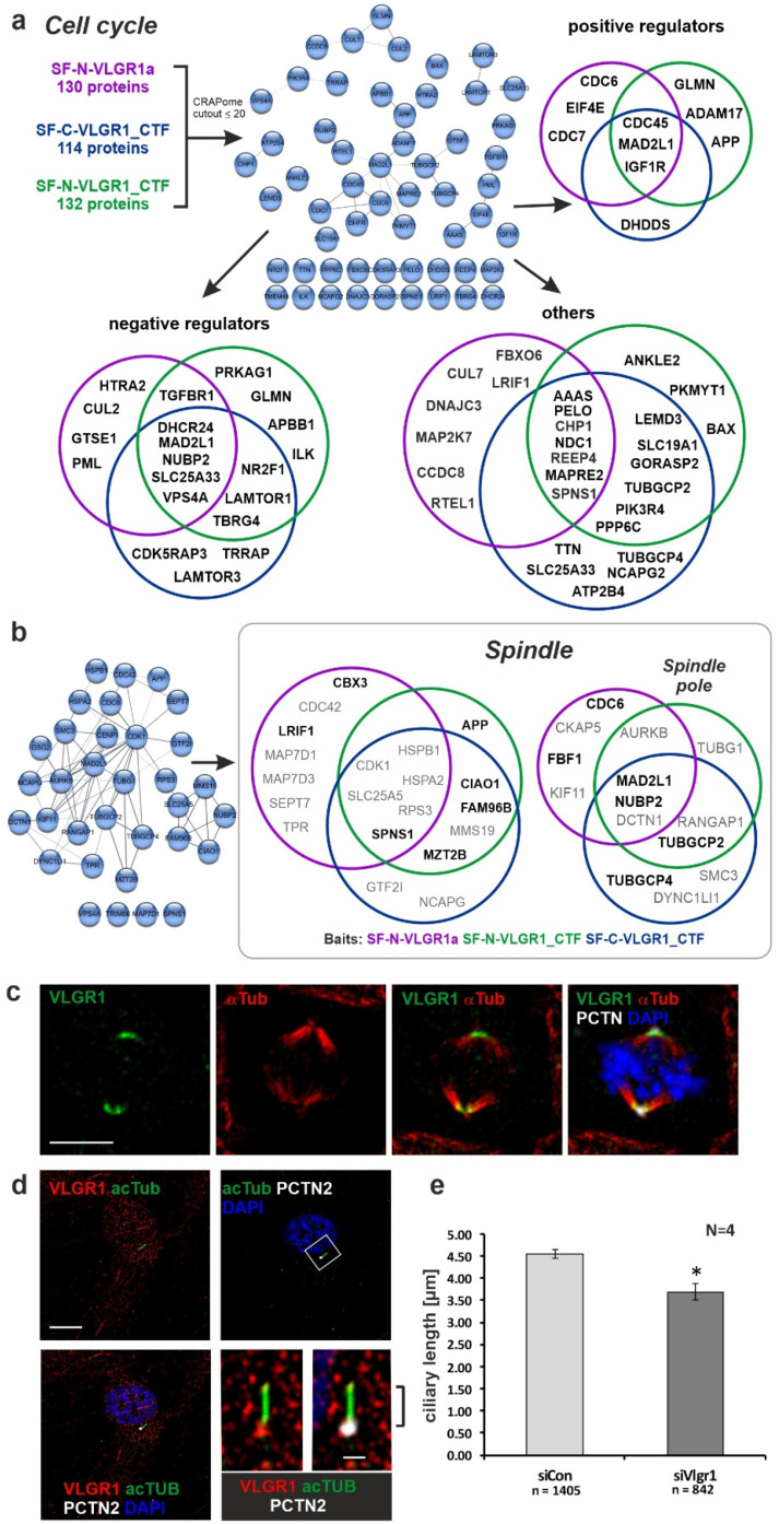
Figure 4.
Association of VLGR1 with cell cycle regulators, mitotic spindle components, and primary cilia. (a) Venn diagrams of VLGR1 prey involved in cell cycle regulation. Interactions between these regulators are visualized in a STRING (https://string-db.org/, accessed on 10 September 2017) network. (b) VLGR1 prey that are assigned to the GO terms spindle and spindle pole in the category Cellular Component. Black font: CRAPome value ≤ 20; Grey font: CRAPome value > 20. (c) Triple immunofluorescence staining of VLGR1 (green), the spindle microtubules by α-tubulin (α-Tub, red), and the centriole/spindle pole marker protein pericentrin-2 (PCTN, white) in RPE1 cells reveals the localization of VLGR1 at spindle poles. Chromosomal DNA is stained by DAPI (blue). (d) Triple immunofluorescence staining revealed VLGR1 (red) co-localization with PCTN2 at the base of primary cilia in RPE1 cells. (e) siRNA-mediated knock-down of VLGR1 in RPE1 cells results in decrease of the ciliary length. Scale bars: 10 µm and 2 µm (magnified primary cilium). siCon, control siRNA (non-targeting); siVlgr1, siRNA directed against VLGR1; * p < 0.05.

