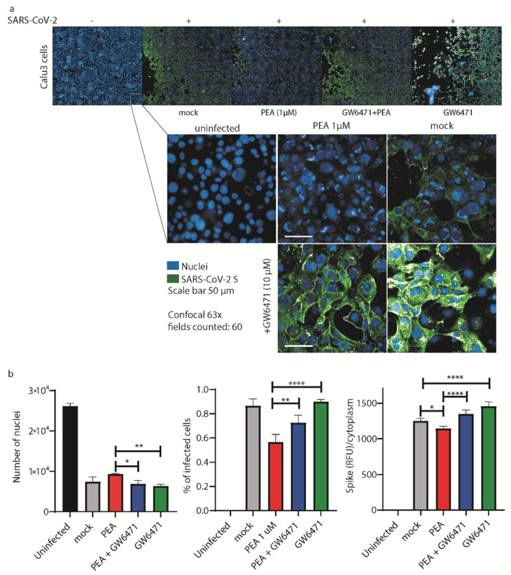
Figure 7.
PEA decreases SARS-CoV-2 entry in Calu-3 cells. (a) (upper panel) High-content automatic confocal microscopy screening of SARS-CoV-2-infected cells (green) treated or not with PEA and PPAR-α antagonist GW6471 (10 μM). (lower panel) Representative images of Calu-3 cells infected or not with SARS-CoV-2 mixed or not with PEA. Cells were also treated or not with GW6471. Sixty fields were acquired per well. (b) Statistical analyses of the number of nuclei detected in the screenings, percentages of infected cells, and the amount of SARS-CoV-2 S protein/cell. Data were analyzed using a 63× water objective. Data are expressed as means ± SD and were analyzed by a one-way ANOVA (n = 3, * p < 0.5, ** p < 0.01, **** p < 0.0001).

