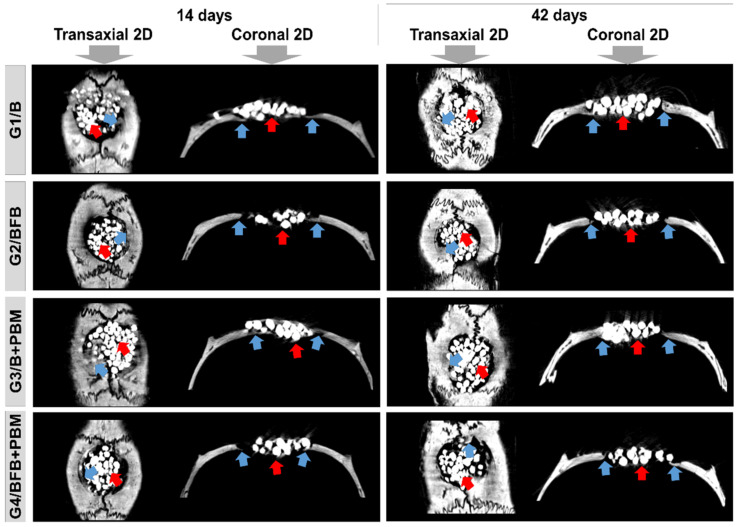
Figure 5.
Two-dimensional (2D) reconstructed microtomographic images in transaxial and coronal sections of the bone defects in rat calvaria at 14 and 42 days, respectively. Defect filled with biomaterial (G1/B), biocomplex consisting of biomaterial plus heterologous fibrin biopolymer (G2/BFB), biomaterial and PBM (G3/B + PBM), and biocomplex consisting of biomaterial plus heterologous fibrin biopolymer and photobiomodulation with low-level laser therapy (G4/BFB + PBM). Bone formation (blue arrow) and biomaterial particles (red arrow).

