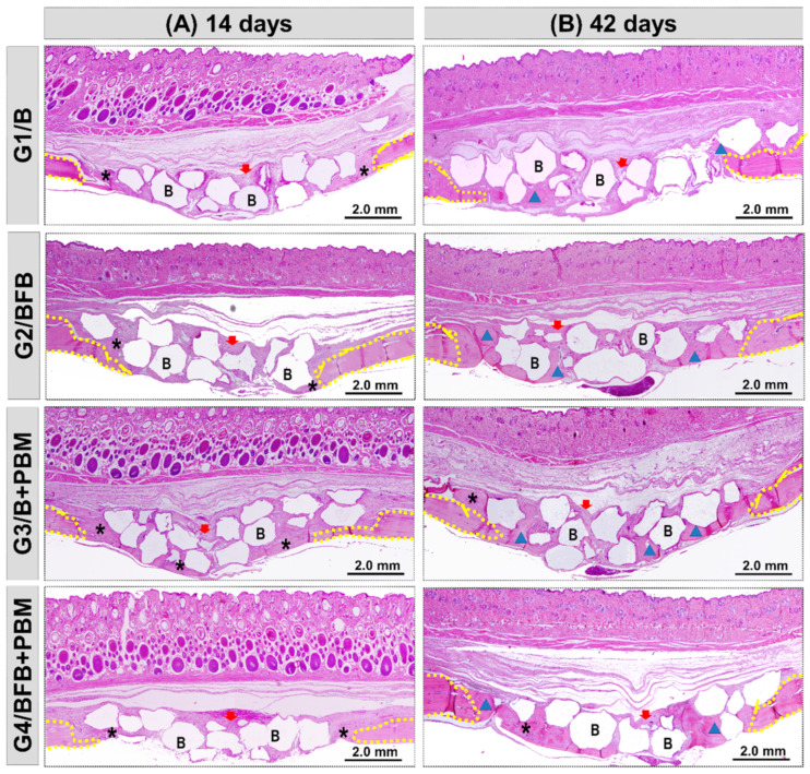
Figure 6.
Panoramic histological views at 14 (A) and 42 (B) days in the cranial defects filled with biomaterial (G1/B), biocomplex consisting of biomaterial plus heterologous fibrin biopolymer (G2/BFB), biomaterial and PBM (G3/B + PBM), and biocomplex consisting of biomaterial plus heterologous fibrin biopolymer and photobiomodulation with low-level laser therapy (G4/BFB + PBM). Immature trabecular formation (asterisk) occurring at the edge of the defect (dashed line) and overlying the dura mater surface. Biomaterial particles (B) permeating the reaction connective tissue (red arrow). The transition from bone maturation to mineralized tissue (triangle), with primary bone areas (asterisk) and biomaterial particles in densely fibrous connective tissue (red arrow). HE; original magnification × 4; bar = 2 mm.

