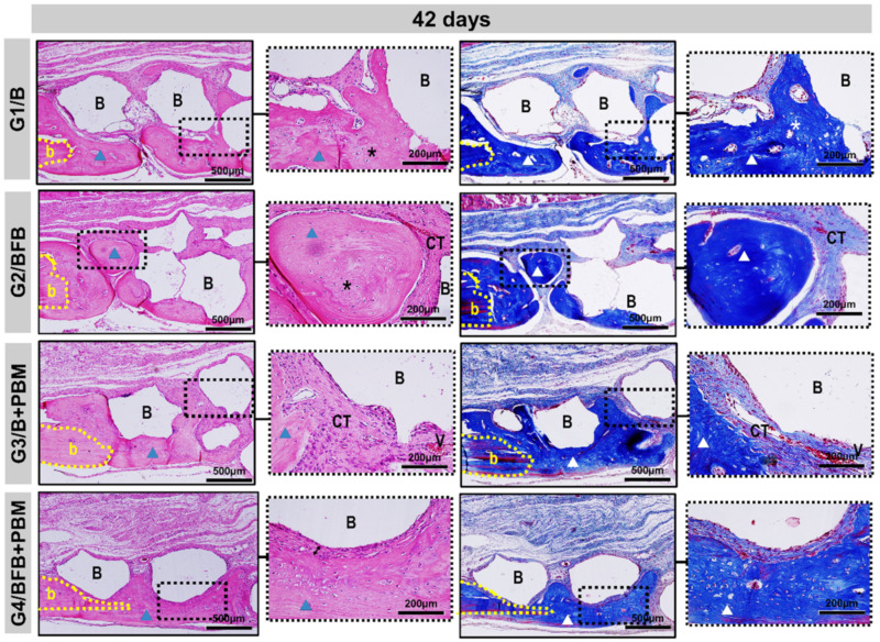
Figure 8.
Details of the evolution of the bone repair process of the cranial defects at 42 days filled with biomaterial (G1/B), biocomplex consisting of biomaterial plus heterologous fibrin biopolymer (G2/BFB), biomaterial and PBM (G3/B + PBM), and biocomplex consisting of biomaterial plus heterologous fibrin biopolymer and photobiomodulation with low-level laser therapy (G4/BFB + PBM). Mature lamellar tissue (triangle) was restricted to the edge of the defect (b) and areas of immature bone trabeculae (asterisk) in the fibrous connective tissue (CT). Biomaterial particles (B) surrounded by thicker collagen fibers, with a fibrous interface between the particles and newly formed bone (arrow). HE and Masson Trichrome; original magnification × 10; bar = 500 µm and insert, images magnified × 40; bar = 100 µm.

