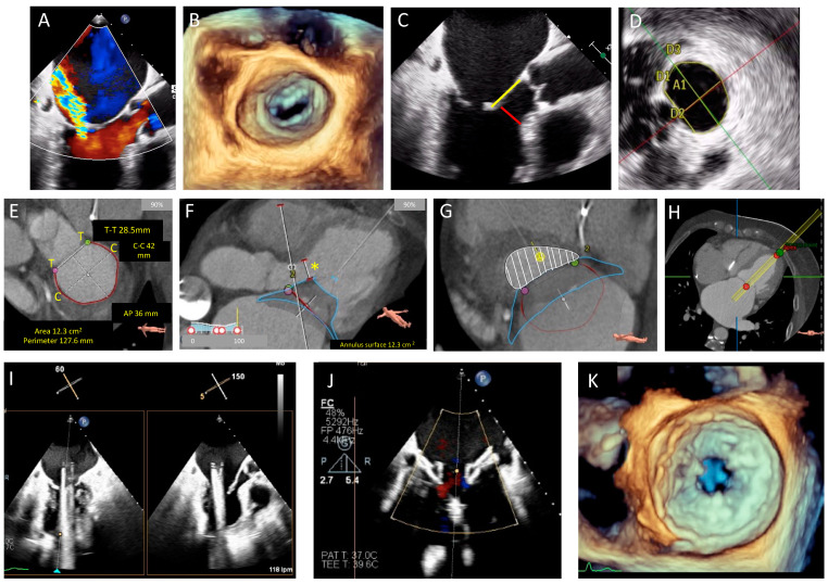
Figure 2.
TEE and cardiac computed tomography (CT) planning for a transcatheter Tendyne™ procedure in a patient with severe MR and calcified leaflets. (A): Severe MR with a posteriorly directed jet, (B): 3D transesophageal echo (TEE) demonstrating calcified leaflet tips unsuitable for edge-to-edge repair. (C): Calcified mitral valve (MV) leaflet tips, anterior MV leaflet (AMVL) length 25 mm (yellow) and AMVL to septum distance of 6 mm (red) (measured to assess risk of left ventricular outflow tract obstruction (LVOTO). (D): 3D assessment of MV annular area (A1 = 12 cm2), perimeter (13.1 cm), AP dimension (D2 = 3.11 cm), inter-trigonal distance (D1 = 3 cm) and intercommisural distance (D3 = 4.6 cm). (E): CT planning demonstrating MV dimensions (T to T = intertrigonal distance, perimeter outlined in red, anteroposterior (AP) distance and intercommisural distance (C to C)). (F): Simulated Tendyne™ with predicted neo-LVOT diameter (yellow asterisk). (G): Simulated Tendyne with predicted neo-left ventricular outflow tract (neo-LVOT) area of 4.6 cm2 (white shaded area), (H): CT assessed apical puncture site for correct orientation with the MV. (I): Intraprocedural TEE guidance with X-plane views showing the delivery system across the MV and in the left atrium (LA), (J): Partial liberation of the Tendyne™ system (LP37M) and assessment of paravalvular leak with colour Doppler. (K): Liberated Tendyne™ valve with no residual MR, no paravalvular leak, mean transvalvular gradient of 5 mmHg, and no dynamic gradient in the LVOT.

