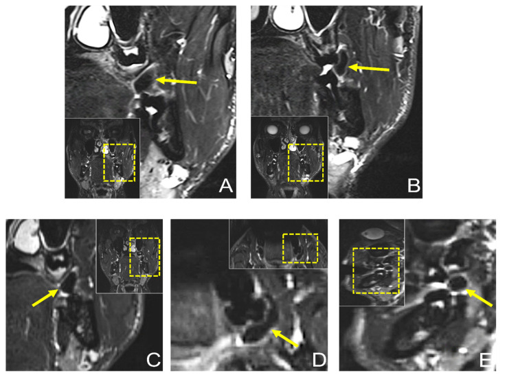
Figure 4.
Preoperative dental MRI showed a well-demarcated lesion with a maximum extension of (D) 21.3 × 5.6 mm (axial), (A–C) 13 × 8.6 mm (coronal), and (E) 10.1 × 9.8 mm (sagittal) on a T2 (short-tau inversion recovery) STIR protocol, with homogenous low signal intensity in the central area of the lesion, while the peripheral area showed a high signal intensity. The lesion originated from the planum buccale and did not infiltrate adjacent structures, with (E) a displacement of the second upper molar. For orientation, the dotted rectangles in the corner show the enlarged area.

