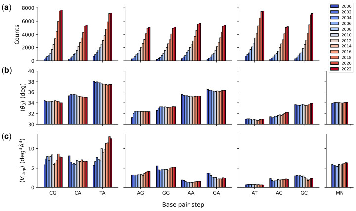
Figure 2.
Color-coded histograms illustrating sequence-dependent features of DNA found within high-resolution protein–DNA structures collected over the last two decades: (a) number of base-pair steps; (b) intrinsic dimer structure measured in terms of the average twist angle between successive base pairs; (c) dimer deformability measured in terms the average volume of configuration space accessed by individual steps. MN parameters for a generic MpN step based on equal weighting of the average parameters of the 16 common dimers. Base-pair steps grouped by chemical class (pyrimidine–purine, purine–purine, and purine–pyrimidine). See Table S2 for numerical values and Methods for details.

