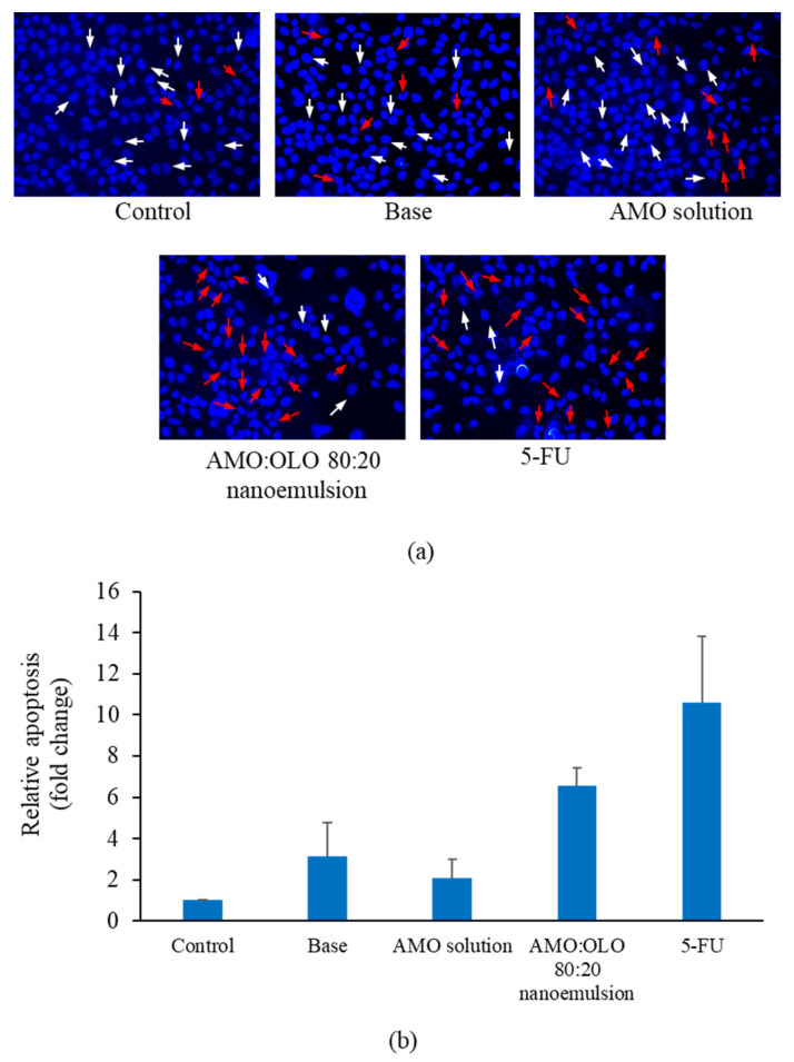
Figure 6.
Nuclear fragmentation of KON cells (a) and relative apoptosis (b) determined by DAPI staining after treatment with control, base (10% w/w of Kolliphor EL), AMO solution (at concentrations equal to those used in nanoemulsions), IC60 of AMO:OLO 80:20 nanoemulsion, and 30 µg/mL of 5-FU. White and red arrows represent normal and apoptotic nuclei, respectively. The values are expressed as mean ± SD (n = 3).

