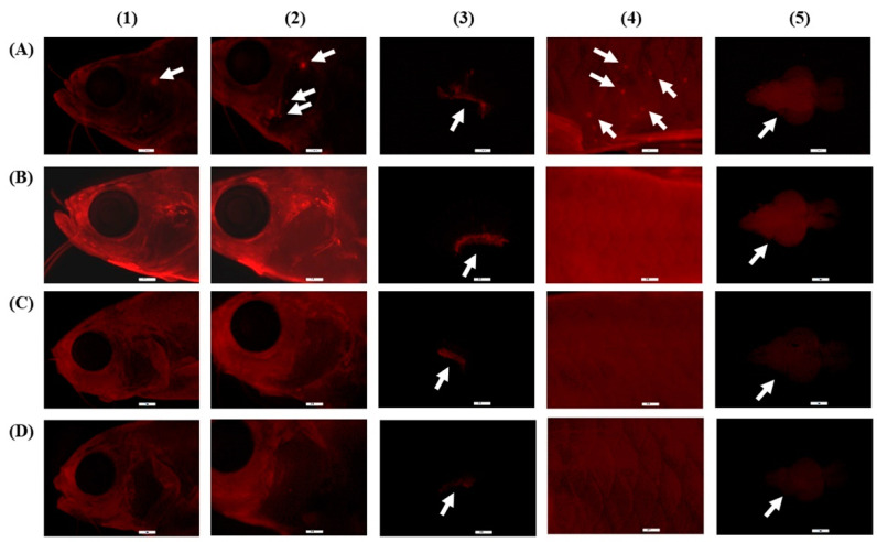
Figure 2.
Fluorescence microscopy images of CONR-EtOH (A), CONR-NE (B), CONR-SMEDDS (C), and CONR-SNEDDS (D) accumulated in external and internal organs of transparent zebrafish: head (1), gill (2), gill filament (3), skin (4), and brain (5). These illustrations are demonstrated for qualitative determination.

