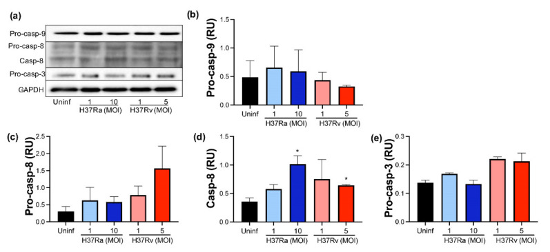
Figure 6.
The presence of virulent M. tuberculosis induces the expression and activation of caspase-8. Human MDMs were obtained after seven days in culture. 2 × 10⁶ MDMs were infected at MOI 1 and MOI 10 with an avirulent (H37Ra) and MOI 1 and MOI 5 with a virulent (H37Rv) strain of M. tb; 2 × 10⁶ MDMs were not infected as a control (Uninf). At 24 h postinfection, cells were recovered and prepared for Western Blot. Representative Western blot of Pro-caspase-9, Pro-caspase-8, Caspase-8, Pro-caspase-3, and GAPDH (a). Band densities of Pro-caspase-9 (b), Pro-caspase-8 (c), Caspase-8 (d), and Pro-caspase-3 (e) were normalized against GAPDH and quantified by densitometry analysis with the ImageJ software. Results are shown in relative units (RU) of concentration. The bar graphs show the mean ± SD from two independent experiments (n = 2 donors and two technical replicates). Statistical analysis was performed using Kruskal–Wallis analysis, followed by Dunn’s post hoc test. * p < 0.05.

