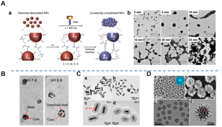
Figure 2.
Morphological design of nanoparticles (NPs). (A). Light-triggered assembly of gold NPs (AuNPs). (Aa) Schematic illustration of a light-triggered assembly of diazirine-decorated AuNPs (dAuNPs). (Ab) Transmission electron microscopy (TEM) images of dAuNPs before and after illumination with a 405 nm laser for different periods of time [42]. Aggregation and the agglomeration degree of dAuNPs depended on irradiation time, demonstrating that interparticle cross-linking took place upon laser irradiation. (B). TEM images of shell-stacked NPs (SNPs) in PBS at pH 7.4 or 6.8 [45]. SNPs with size and charge dual-transformable ability displayed a clear spherical core–shell structure at pH 7.4, with a size of 145 nm. When SNPs were incubated at pH 6.8, a polyethylene glycol (PEG) corona detached from the core and subsequently the small-sized core with a size of 40 nm was exposed. (C). Morphology and structure of gold nanorods (GNRs) and bacteria-like mesoporous silica nanoshell (MSN)-coated GNRs (bGNR@MSN). (Ca) TEM image of GNRs. (Cb,Cc) TEM images of bGNR@MSN coated for 12 h with silica. The red arrows indicate the size (~6.7 nm) of mesopores. (Cd) TEM image of bGNR@MSN coated for 24 h with silica [29]. The morphology of the outside mesoporous silica layer resembled bacterial pili, and the thickness of the mesoporous silica layer could be controlled by changing the reaction time. (D). Morphology of virus-like mesoporous silica NPs. (Da,Db) Scanning electron microscopy (SEM) and (Dc,Dd) TEM images with different magnifications of the virus-like mesoporous silica NPs. The red arrows mark the open tubular structures; the red circles highlight the top view of the open silica nanotubes. The inset of (Da) is a structural model for the virus-like mesoporous silica [47]. (Image (A) is reproduced with permission from [42] (Copyright © 2016 John Wiley & Sons, Inc.). Image (B) is reproduced with permission from [45] (Copyright © 2017 John Wiley & Sons, Inc.). Image (C) is reprinted with permission from [29] (Copyright © 2018 Elsevier Ltd.). Image (D) is reprinted with permission from [47] (Copyright © 2017 American Chemical Society).)

