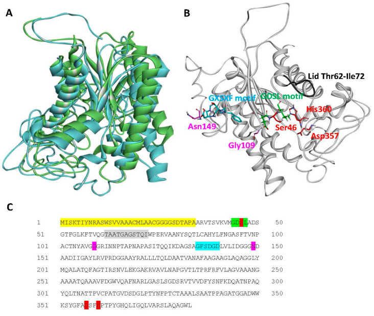
Figure 3.
Structural analysis of A. citrulli M6 Lip1. (A) Structural alignment between Lip1 (blue ribbon), and the passenger domain of the autotransporter EstA from Pseudomonas aeruginosa (green ribbon; PDB code 3KVN). (B) Predicted structural model of Lip1 generated by I-TASSER, and (C) Lip1 sequence. Conserved residues and motifs are highlighted and shown in sticks as follows: red, catalytic triad; green (and red for Ser46), GDSL motif; blue, GXSXG motif; purple, oxyanion hole residues; and grey, putative lid loop (ribbon only). The secretion signal peptide as predicted by Pred-Tat and Phobius is highlighted yellow in the protein sequence.

