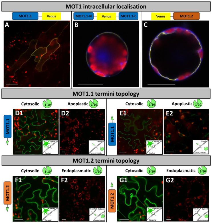
Figure 1.
Intracellularlocalisation of MOT1 family members and termini topology. Localisation studies of MOT1 family members (A–C) were performed by transient transformation of (A) A. thaliana via FAST method, and (B,C) N. benthamiana mesophyll protoplasts via chemical transformation to express fusion proteins of Venus and MOT1.1 in different orientations. N. benthamiana protoplasts were co-transformed with cytosolic marker eqFP611. Images were merged from (A) Venus, chlorophyll auto-fluorescence channels, and also (B,C) with the eqFP611 fluorescence channel. Split-GFP topology studies (D–G) were carried out with transient transformation by Agrobacterium infiltration of N. benthamiana leaves to express GFP11-MOT fusion proteins. Two days later, leaves were co-transformed with GFP1-10 (D1,E1,F1,G1); SP-GFP1-10 (D2,E2); and SP-GFP1-10-HDEL (F2,G2). Images were taken 2–3 days after transformation and merged from GFP and chlorophyll auto-fluorescence channels. Each image was taken with a C-Apochromat 40×/1.2 water immersion objective and scale bars depict a length of 20 μm.

