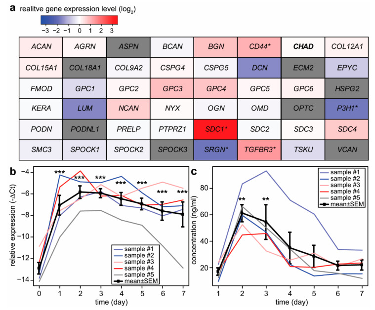
Figure 2.
Changes in proteoglycan gene expression during villous trophoblast differentiation. (a) Microarray data were obtained from primary villous trophoblast cells isolated from third-trimester normal placentas (n = 3) during a seven-day differentiation period. The largest differences in gene expression compared to day 0 were visualized on a heatmap. Color code depicts log2 gene expression ratios. Grey color: no data were available. The original study was published by Szilagyi et al. [52]. (b) SDC1 expression was monitored during villous trophoblast differentiation. qRT-PCR data were obtained from an extended set of primary villous trophoblast cells isolated from third-trimester normal placentas (n = 5) during a seven-day differentiation period. Relative expression of SDC1, normalized to RPLP0, was visualized on the diagraph. (c) Changes in syndecan-1 protein concentration in cell culture supernatants (n = 5) were examined throughout spontaneous syncytial differentiation of primary villous trophoblast cells. One-Way ANOVA with Dunnett’s post-hoc test was used for the analysis of qRT-PCR and ELISA results (* p < 0.05, ** p < 0.01, *** p < 0.001). Ribosomal Protein Lateral Stalk Subunit P0—RPLP0; syndecan-1—SDC1.

