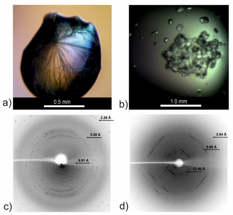
Figure 8.
Crystallization drops and X-ray diffraction images for the CysZ9 peptide crystallized in (a) needle-shaped form from 2.4 M sodium malonate pH 6.0, and in (b) cuboid form from 0.2 M MgCl2, 10% MPD, 0.05 M sodium cacodylate pH 6.5, 0.001 M spermine. (c) Sample diffraction image recorded for crystal from (a); (d) sample diffraction image recorded for crystal form (b).

